Figure 2.
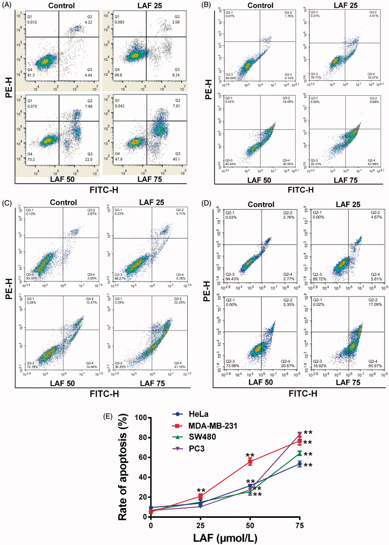
Lappaol F (LAF) promoted apoptosis in cancer cells. LAF (0, 25, 50 or 75 µmol/L) was incubated with cancer cells for 48 h. The rate of apoptosis was analysed by flow cytometry after annexin V-FITC/PI staining. (A) Representative plots of HeLa cells. (B) Representative plots of MDA-MB-231 cells. (C) Representative plots of SW480 cells. (D) Representative plots of PC3 cells. (E) Quantification of apoptosis (including early and late apoptotic cells). All data are expressed as the mean ± SD (n = 3). **p < 0.01, significantly different from the control without LAF treatment.

