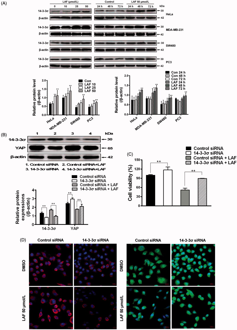
Figure 6.
Lappaol F (LAF) inhibited YAP by increasing 14-3-3σ levels. (A) HeLa, MDA-MB-231, SW480 and PC3 cells were treated with LAF at different concentrations for 48 h (left side), or at 50 µmol/L LAF for different durations (right side). Western blotting was performed to evaluate the levels of 14-3-3σ. (B-D), HeLa cells were transfected with 14-3-3σ siRNA followed by LAF (50 µmol/L) treatment for 24 h. (B) The protein levels of 14-3-3σ and YAP were measured by western blotting. (C) Cell viabilities were measured by sulforhodamine B assay. (D) The expression and location of 14-3-3σ (red color) and YAP (green color) were observed by immunofluorescence staining. Nuclei (blue color) were stained with 4,6-diamino-2-phenyl indole. Scale bars represent 20,000 nm. All data are expressed as the mean ± SD (western blotting and immunofluorescence, n = 3; cell viability, n = 6). *p < 0.05, **p < 0.01, significantly different from the control siRNA group. DMSO: dimethyl sulfoxide; siRNA: small interfering RNA.

