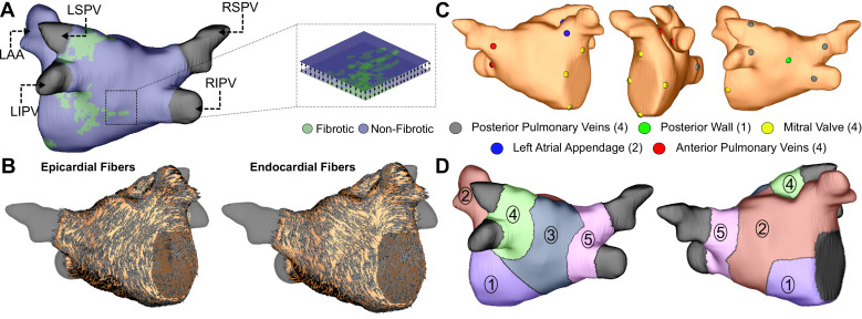
Figure 1. Model generation.
(A) Reconstruction of LA geometry with anatomical features labeled (RIPV/RSPV/LIPV/LSPV, right/left inferior/superior pulmonary veins; LAA, LA appendage). The LA is modeled as a bilayer comprising nested endocardial and epicardial shells linked in both fibrotic and non-fibrotic regions by 1D linear elements. (B) LA fiber orientations for the endocardium and epicardium, mapped from human atlas geometry as described in Materials and methods. (C) AFib trigger sites as pacing sites (posterior/anterior LIPV, LSPV, RSPV, RIPV, LAA base, mitral valve annulus, and posterior wall). (D) Regions of the LA generated as described in methods: (Andrade et al., 2014) atrial floor, (Israel et al., 2017) anterior wall and LAA, (Brachmann et al., 2016) posterior wall, (Tandon et al., 2019) left PVs, and (Zahid et al., 2016a) right PVs.


