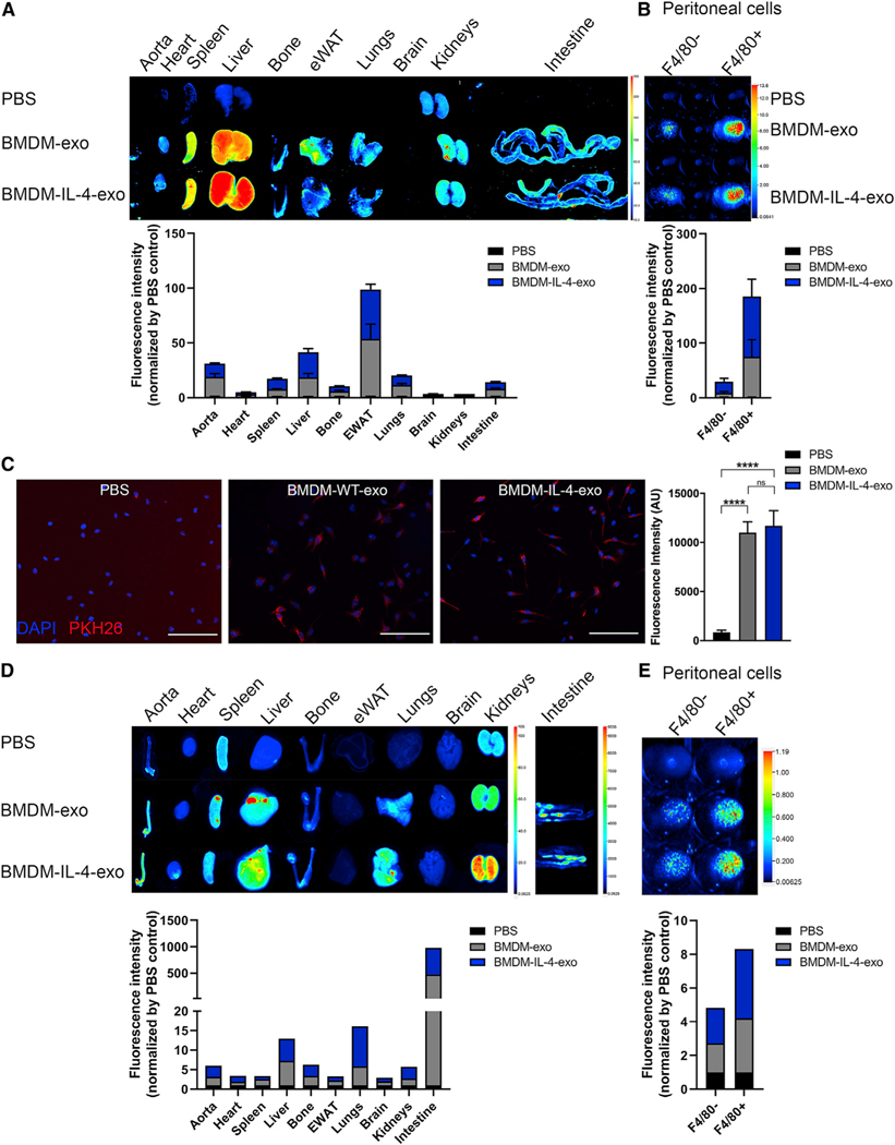
Figure 2. Biodistribution of Macrophage Exosomes and Delivery of Their microRNA Cargo to Tissues of Apoe−/− Mice.
Exosomes purified from BMDM culture medium were labeled with fluorescent lipophilic tracer DiR and infused into 25-week-old Apoe−/− mice for 4 weeks every 2 days.
(A) Representative images of organs (12 h post-injection) from mice injected i.p. with 1 × 10¹⁰ particles (measured using NTA) and quantification using the PBS as background control.
(B) Representative images of peritoneal F4/80− and F4/80+ isolated cells from mice injected with DiR-labeled exosomes. Fluorescence of DiR-labeled BMDM exosomes was detected using the LI-COR Odyssey infrared imaging system. Color scale indicates fluorescence intensity.
(C) Merged images showing internalization of PKH26-labeled BMDM-exo (red) by naive culture BMDMs counterstained with DAPI (blue). BMDMs were co-incubated with 2 × 10⁹ PKH26-labeled exosomes for 2 h at 37°C and washed repeatedly to remove unbound exosomes. All images were acquired using a Zeiss microscope system with a 20× objectives. Scale bars: 100 μm. Statistical analysis of fluorescence intensity was conducted using one-way ANOVA and Tukey’s multiple-comparisons test.
(D and E) Distribution of synthetic IRDye-labeled miR-146b, transfected into BMDM exosomes, 2 h after i.p. injection in 25-week-old Apoe−/− mice (D). Representative images of organs and (E) peritoneal F4/80− and F4/80+ isolated cells from mice injected with IRDye-labeled miR-146b-loaded exosomes detected using the LI-COR Odyssey infrared imaging system.****p < 0.0001 as determined using one-way ANOVA and Tukey’s post-hoc test. Data are represented as mean ± SEM.

