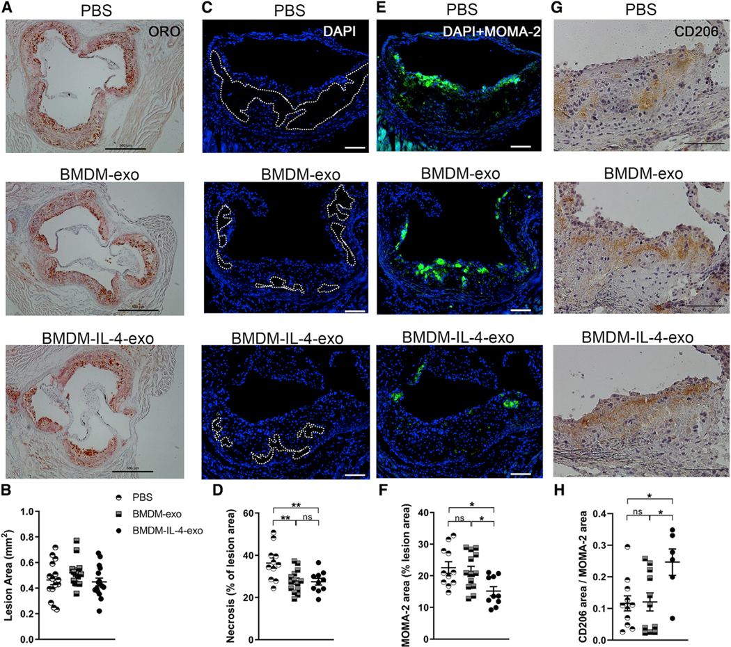
Figure 5. Resolution of Inflammation in Atheroma of Apoe−/− Mice Treated with BMDM-IL-4-Exo.
(A and B) Histological analysis (A) and (B) quantification of cross sections of the aortic sinus stained with oil red O (ORO) from 25-week-old Apoe−/− mice fed with a Western diet and injected with PBS, BMDM-exo, or BMDM-IL-4-exo for 4 weeks. Scale bar, 500 μm. n = 13–15 in each group.
(C) Representative cross-sectional view of aortic root stained with DAPI to measure necrosis area from each group of mice. Dashed lines show the boundary of the developing necrotic core. Scale bar, 100 μm.
(D) Quantification of necrotic core area as a percentage of total plaque area.
(E and F) Representative images (E) and (F) quantification of MOMA-2+ macrophages in the atherosclerotic plaques of aortic root areas. Scale bar, 100 μm.
(G) Representative image of CD206 staining in aortic root lesions. Scale bar, 100 μm.
(H) Quantification of CD206 staining as a ratio of macrophage lesion area. Results from a pool of three independent experiments are shown; n = 11–14 in each group.
Statistical analysis was performed using one-way ANOVA and Sidak’s multiple-comparisons post-test. *p < 0.05 and **p < 0.01. Data are represented as mean ± SEM.

