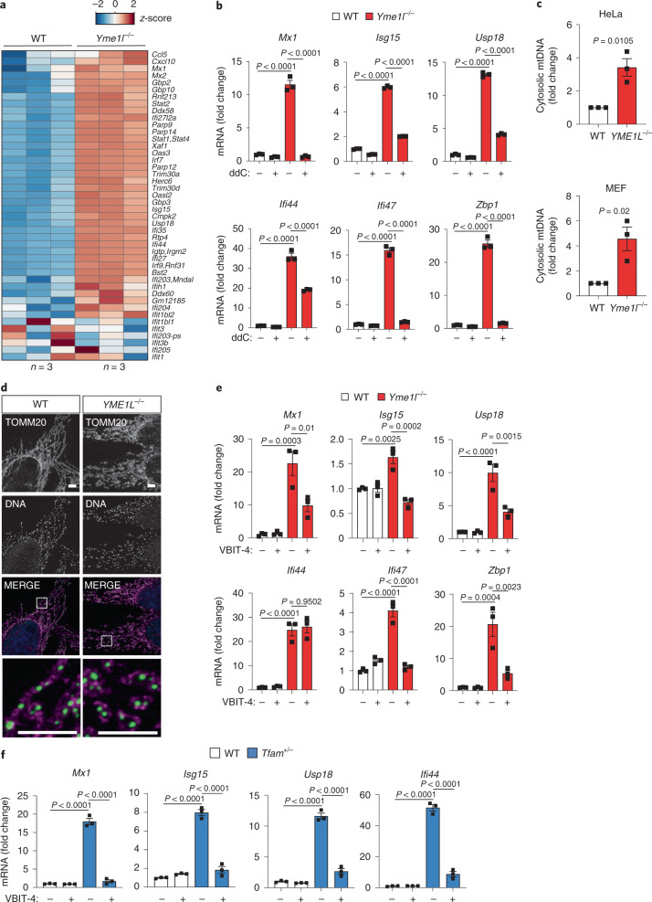
Fig. 2. mtDNA is released into the cytosol in YME1L-deficient cells.
a, Expression of ISGs, which were previously described as being induced by mtDNA stress upon depletion of TFAM (including in addition Mx1)4,5, in WT versus Yme1l−/− MEFs (RNA-Seq; z-score normalized log2(FPKM) values, n = 3 independent cultures). b, ISG expression in MEFs treated with water or ddC (20 µM) for 9 days (n = 3 independent cultures). c, mtDNA levels in cytosolic fractions from HeLa cells and MEFs assessed by qPCR amplification of mitochondrial CYTB (n = 3 independent cultures). d, Immunocytochemistry of HeLa cells using antibodies against TOMM20 (mitochondria) and DNA, scale bar, 5 µm (n = 3 independent cultures). e, ISG expression in WT and Yme1l−/− MEFs treated with 10 µM of the VDAC1 oligomerization inhibitor VBIT-4 for 48 h (n = 3 independent cultures). f, ISG expression in WT and Tfam−/− MEFs treated with 10 µM VBIT-4 for 48 h (n = 3 independent cultures). P values calculated using two-way ANOVA with Tukey’s multiple comparison test (b,e,f) or two-tailed unpaired t-test (c). Data are means ± s.e.m.

