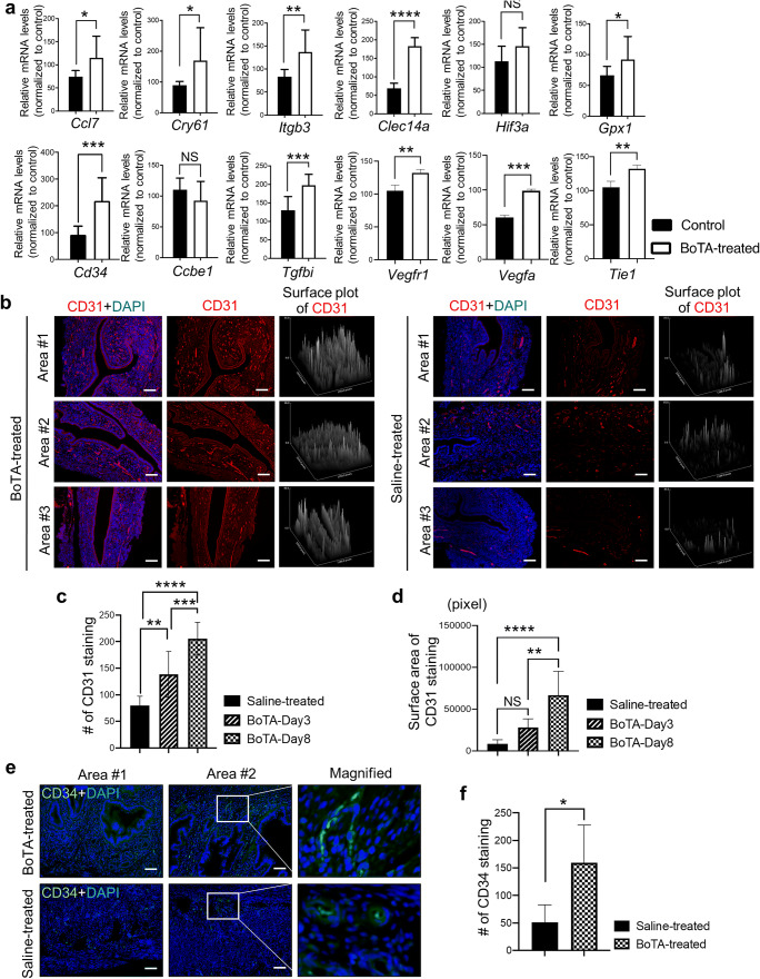
Fig. 4.
Induction of angiogenesis-related genes in BoTA-administered mouse uterus. (a) QRT-PCR analysis of Ccl7, Cry61, Itgb3, Clec14a, Hif3a, Gpx1, Cd34, Ccbe1, Tgfbi, Vegfr1, Vegfa, and Tie1 in BoTA-treated uterus samples compared to controls. (b) IF staining of CD31 in longitudinally sectioned mouse uterus harvested 8 days after BoTA intrauterine infusion. Saline-treated endometrium was used for control. Scale bar: 100um. Representative two images from the different areas are shown in Supplementary Figure 4A. CD31 expression was quantified by counting the number of CD31 staining (c) and stained surface area (d). (e) IF staining of CD34 in longitudinally sectioned mouse uterus harvested 8 days after BoTA intrauterine infusion. Saline-treated endometrium was used for control. Scale bar: 100um. CD34 expression was quantified by counting the number of CD34 staining (f). Comparison groups for (a) and (f) are from 3 independent experiments and analyzed using the unpaired Student t-test for parametric distributions and the multiple comparisons for (c) and (d) are from 3 independent experiments and analyzed using the ordinary one-way ANOVA analysis with Dunnett’s multiple comparison test including P-values (*<0.05, **<0.01, ***<0.001, ****<0.0001, NS not significant)

