Fig. 5.
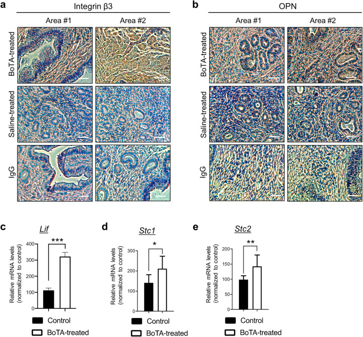
Induction of endometrial receptivity-related genes in BoTA-administered mouse uterus. Representative two images of immunohistochemistry analyses of integrin β3 (a) and OPN (b) in longitudinally sectioned mouse uterus harvested 8 days after BoTA intrauterine infusion. Saline-treated endometrium was used for control. For the negative control, mouse or rabbit IgG was used. Scale bar: 20um. QRT-PCR analysis of Lif (c), Stc1 (d), and Stc2 (e) in BoTA-treated uterus samples compared to controls. Comparison groups for (c–e) are from 3 independent experiments and analyzed using the unpaired Student t-test for parametric distributions including P-values (*<0.05, **<0.01, ***<0.001)

