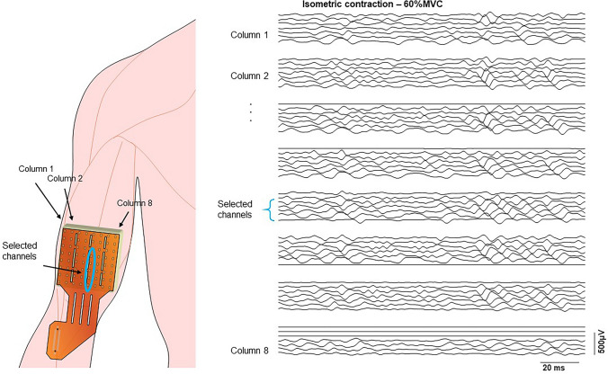
Fig. 1.
Representation of the position of sEMG array on the biceps brachii muscle. An example of EMG signals detected in single differential mode from each column of a FSHD patient during an isometric elbow flexion at 60% MVC is shown on the right panel. The innervation zone can be identified by the V shape of the signals. The selected channels for muscle fiber estimation are located in the distal portion of column 5, where the pure propagation of motor unit action potentials is visible between the innervation zone and the distal tendon

