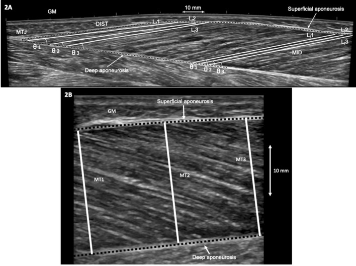
Fig. 2.
Sagittal plane ultrasound images showing examples of muscle architecture measurements of the gastrocnemius medialis (GM) muscle. a GM extended field-of-view image with fascicle length (Lf) and fascicle angle (θ) determination at middle (MID) and distal (DIST) portions of the muscle. For each site, three fascicles (Lf1, Lf2, and Lf3) and their respective angles (θ1, θ2, and θ3) were identified and measured. MTJ muscle–tendon junction. b GM single snapshot in MID showing the three sites at which muscle thickness (MT) was measured (MT1, MT2, and MT3) as the perpendicular distance between the superficial and deep aponeuroses

