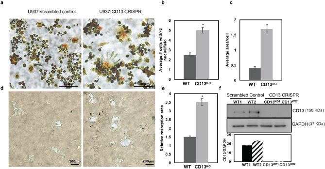
Figure 5.
Increased Multinucleated OC formation in U937 cells expressing CD13 CRISPR. (a). TRAP staining of U937 expressing scrambled control or CD13 CRISPR grown in presence of PMA for 3 days followed by M-CSF and RANKL for 10 d indicated increased size and number of multinucleated OCs in absence of CD13 compared to WT. (b) Multinucleated OC (**) with greater than 3 nuclei per cell and (c), average area of OC. (d). Phase contrast imaging of area of resorption in U937 cells expressing CD13 CRISPR or scrambled control grown in presence of PMA followed by M-CSF and RANKL for 17 d on osteoplates. TRAP+ osteoclasts were imaged with Zeiss fluorescence inverted microscope and analyzed by using Zeiss Zen 2.0 Pro blue edition software (https://www.zeiss.com/content/dam/Microscopy/Downloads/Pdf/FAQs/zen2-blue-edition_installation-guide.pdf). Area of resorption was imaged using a light microscope (Olympus Scientific), using Olympus cellSens Dimension V0118 software (Olympus Scientific) (https://www.olympus-lifescience.com/en/software/cellsens/) quantified by Image J (https://imagej.nih.gov/ij/) (e). (f). Immunoblot analysis of CD13 expression in U937 cells expressing CD13 CRISPR or scrambled control clones. Blots were imaged by ChemiDoc Imaging system version 3.0.1 (https://www.bio-rad.com/en-us/category/chemidoc-imaging-systems?ID=NINJ0Z15) (Biorad). A cropped image is shown, see Supplementary Fig. S6 for full-length blots and cropped replicates. Scale bar-(a) 100 µm; (d) 200 µm. Data represents ± SEM of three independent experiments. N = 3/genotype, *p < 0.05. Magnification ×10.

