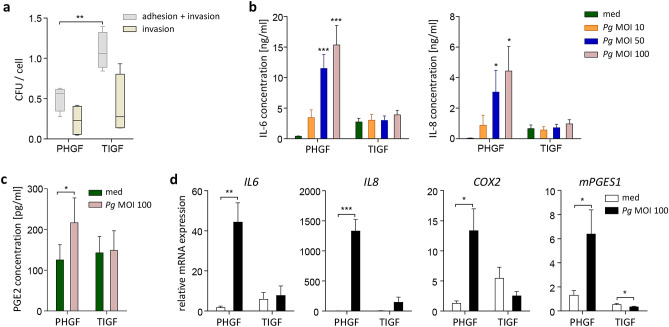
Figure 1.
TIGFs internalize P. gingivalis but fail to mount an inflammatory response after bacterial challenge. (a) P. gingivalis internalization by PHGFs and TIGFs determined by colony-forming assay. Cells were infected with P. gingivalis (MOI 100) for 1 h and then cultured for another 1 h in medium, with or without antibiotics. Results of four independent experiments are presented as boxplots, where the line within the box denotes the median number of colony-forming units (CFU)/cell, the boxes represent the 25th and 75th percentiles, and the lines outside the box mark the minimum and maximum values. **P < 0.01; unpaired t-test (b) Secretion of IL-6 and IL-8 by PHGFs and TIGFs (n = 6–7) infected with increasing MOI of P. gingivalis (10, 50, 100) for 1 h followed by washing, then 23 h culture in fresh medium. *P < 0.05; ***P < 0.001; 1-way ANOVA followed by Bonferroni multiple comparison test. (c) Production of PGE2 by PHGFs and TIGFs (n = 8–9) infected with P. gingivalis (MOI 100) for 24 h. *P < 0.05; paired t-test. Data in (b,c) are presented as mean concentration + SEM. (d) Relative mRNA expression of IL6, IL8, COX2 and mPGES1 in PHGFs and TIGFs (n = 5–7) infected with P. gingivalis (MOI 100) for 24 h. Data are presented as mean relative expression + SEM. *P < 0.05; **P < 0.01; ***P < 0.001; paired t-test.

