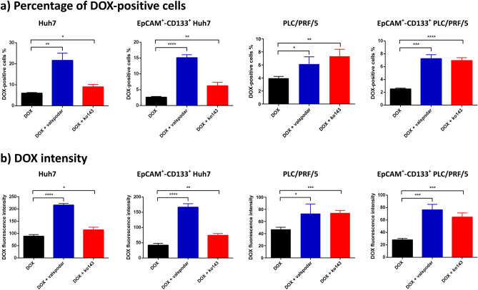
Figure 3.
Involvement of MDR1 and ABCG2 in doxorubicin (DOX) efflux as determined by pharmacological inhibitors. The Huh7 or PLC/PRF/5 cells were treated with 200 nM or 100 nM of DOX in the presence or absence of MDR1 inhibitor valspodar (1 µM) or ABCG2 inhibitor ko143 (1 µM) for 24 h followed by flow cytometric analysis. (a) Percentage of DOX-positive cells; (b) DOX intensity followed by indicated treatments. Data shown are means ± SD, (n = 3). ****p < 0.0001; ***p < 0.001; **p < 0.01; *p < 0.05; compared with DOX treatment.

