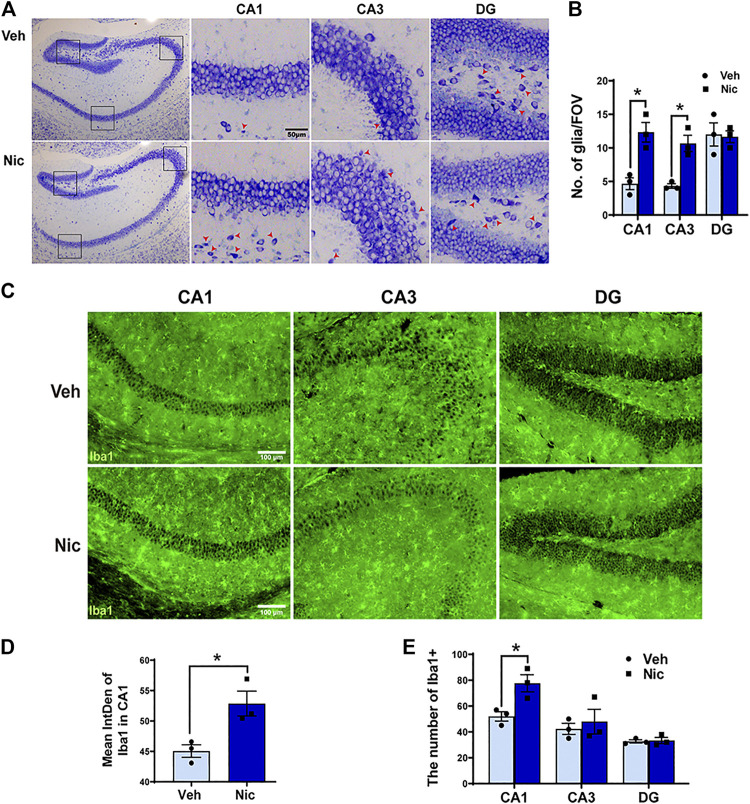
FIGURE 2.
Maternal nicotine exposure increased the number of neuroglia microglia, but not pyramidal neurons in the hippocampus of pups. (A) Photomicrograph showed the whole hippocampal sample in the coronal plane (two pictures on the left-most side). DG, CA1, and CA3 subfield are indicated by the black frame in the left-most image. Maternal nicotine exposure increased neuroglia (arrowhead) in DG, CA1, and CA3. (B) Neuroglial cells were quantified in the hippocampus. (C) Iba1-positive cells in the offspring’ hippocampi were analyzed by immunofluorescence staining using floated section. The hippocampus of 20 day old offspring from the maternal nicotine-exposed group has more microglia cells (green) than the vehicle control. Representative images were shown. The quantification of immunofluorescence staining by ImageJ showed that the expression (D) and number (E) of Iba1 + cells increased obviously in the nicotine group. Scale bar: 50 μm (A); 100 μm (B); *p < 0.05, all data are means ± SEM.

