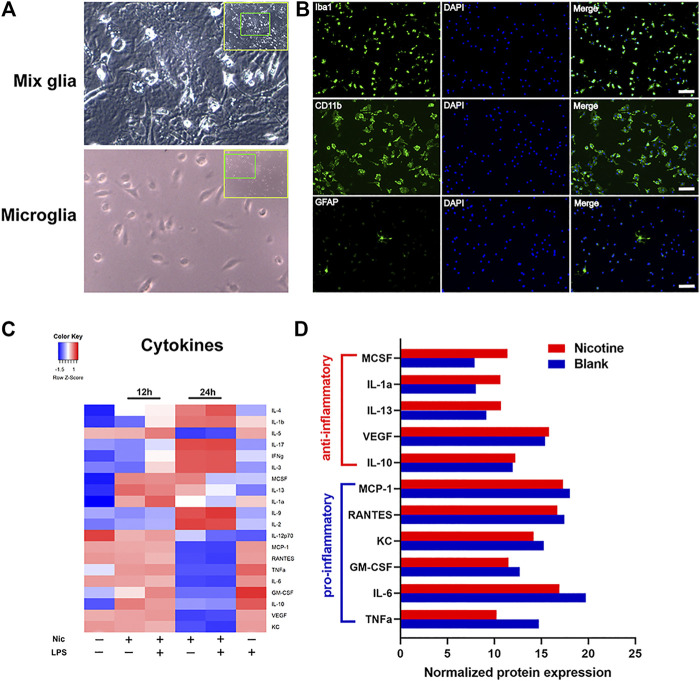
FIGURE 4.
Nicotine exposure suppressed inflammatory cytokine revealed by protein array. Primary microglia were isolated from postnatal day 2–4 mouse brain (A) and were identified by immunofluorescent staining with Iba1, CD11b, and GFAP antibody. Purified microglia were shown in (B). GFAP positive astrocytes were found scarcely in the culture while the vast majority of cells were positive for Iba1 and CD11b staining. Unstimulated primary microglia were served as blank for each time point. LPS (10 μg/ml)-stimulated cells (LPS group), nicotine (10 μmol)-treated cells (nicotine group), and LPS (10 μg/ml) plus nicotine (10 μmol)-treated cells (LPS plus nicotine group) were used. Cells were pretreated with LPS 30 min before adding nicotine. Microglia cells incubated with LPS for 24 h were used as the pro-inflammatory control. Supernatant were collected at 12 and 24 h after treatment. Each group had duplicated wells. Pro-inflammatory and anti-inflammatory responses in microglia were evaluated by a commercial cytokine array. Cytokines expression levels were severally determined using a 20-array. Heat map representing cytokine concentrations was shown (C). Quantitative analysis of cytokines secreted from primary microglia were presented in the graph (D). Each group was duplicated, and the mean of the respective cytokine is represented in the heat map and the bar graph.

