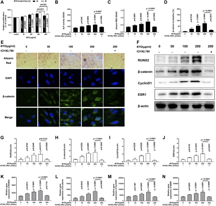
FIGURE 4.
BYD activated Wnt/β-catenin signaling pathway to the promoted osteoblastic formation in vitro via ESR1. (A) Relative proliferation of various concentration of BYD (50, 100, 200, 400 μg/ml). (B) Alkaline phosphatase (ALP) activity. (C) Result of Alizarin Red staining. (D) β-catenin localization analysis result. (E) Representative images of alizarin red staining and immunofluorescence staining of β-catenin. (F) Representative images of western blots of RUNX2, β-catenin, cylindD1, and ESR1. Quantitative analysis results of protein expressions analysis of (G) RUNX2, (H) β-catenin, (I) CyclinD1, and (J) ESR1. Quantitative analysis results of mRNA expression analysis of (K) RUNX2, (L) β-catenin, (M) CyclinD1, and (N) ESR1. THE exact p-value can be found in the corresponding histogram.

