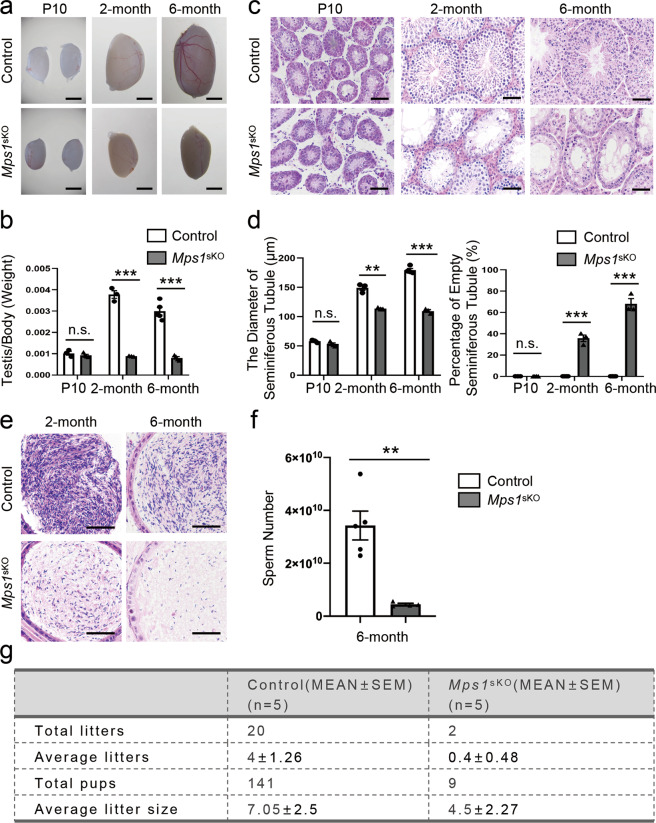
Fig. 2. Postnatal disruption of Mps1 led to severe defects in spermatogenesis and significantly reduced male fertility.
a Image of control and Mps1sKO testes at P10, 2 months of age, and 6 months of age. Scale bar = 2 mm. b Ratios of testis weight to body weight for control and Mps1sKO mice at P10, 2 months of age, and 6 months of age. n ≥ 3; Student’s t test; n.s. no significance; ***P < 0.001. c H&E staining of control and Mps1sKO testes at P10, 2 months of age and 6 months of age. Scale bar = 50 µm. d Statistical results for the seminiferous tubule diameters and empty seminiferous tubule percentages of control and Mps1sKO testes at P10, 2 months of age, and 6 months of age. At least 100 tubules were counted from at least 3 different mice; Student’s t test; **P < 0.01, ***P < 0.001. e H&E staining of control and Mps1sKO cauda epididymides from 2 and 6-month-old mice. Scale bar = 50 µm. f Sperm counts from the cauda epididymides of at least four different mice at 6-month of age. Student’s t test; **P < 0.01. g Fertility test of male control and Mps1sKO mice. In each group, five 3-month-old male mice were individually mated with two wild-type female mice for 3 continuous months.

