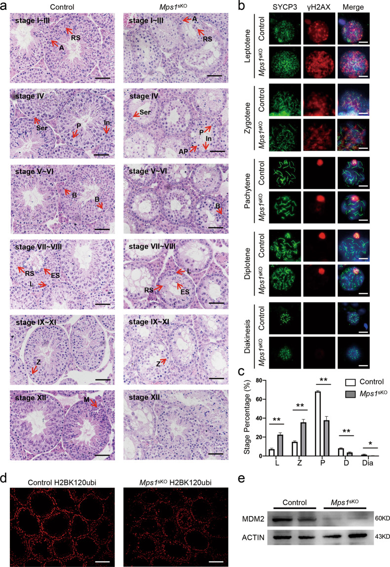
Fig. 5. In Mps1sKO mouse testes, the zygotene-to-pachytene transition was delayed, and the progression of meiosis I was compromised.
a Representative H&E staining of control and Mps1sKO seminiferous tubules. A type A spermatogonia, In intermediate spermatogonia, B type B spermatogonia, P pachytene spermatocytes, L leptotene spermatocytes, Z zygotene spermatocytes, RS round spermatids, ES elongated spermatids, M meiotic divisions, AP apoptosis pachytene, Ser Sertoli. Scale bar = 50 µm. b Meiotic spermatocyte spreads for control and Mps1sKO mice at 1-month of age. Co-staining was performed with SYCP3 and γH2AX antibodies. Scale bar = 10 µm. c Percentage of meiotic spermatocytes in each stage. L leptotene, Z zygotene, P pachytene, D diplotene, Dia diakinesis. A total of 650–1000 spermatocytes were counted from 3 different mice. Student’s t test; *P < 0.05, **P < 0.01. d Immunofluorescence staining of H2BK120ubi in control and Mps1sKO testes at 2 months of age. Scale bar = 100 µm. e Western blot of MDM2 in control and Mps1sKO testes at 2 months of age.

