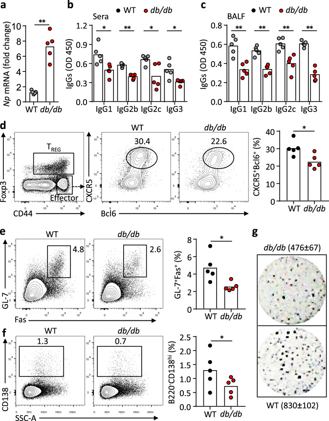
Fig. 3. LepR deficiency leads to impaired antibody responses after influenza virus infection.
a Real-time PCR analysis of nucleoprotein (Np) in lung tissues of WT (grey) and db/db (red) mice 9 days post H1N1 influenza viral infection (**P = 0.0007). b, c ELISA measurement of H1N1-specific IgG1, IgG2b, IgG2c and IgG3 in sera (b) (IgG1: *P = 0.0190, IgG2b: **P < 0.0001, IgG2c: *P = 0.0299, IgG3: *P = 0.0209) and BALF (c) (IgG1: **P = 0.0080, IgG2b: **P = 0.0036, IgG2c: **P = 0.0079, IgG3: **P = 0.0007). d–f Representative FACS plots and statistics showing CXCR5+Bcl6+ TFH cells (d) (*P = 0.0407), GL-7+Fas+ germinal centre (GC) B (e) (*P = 0.0240) and CD138high antibody-secreting cells (ASCs) (f) (*P = 0.0411) in mediastinal lymph nodes. g ELISPOT assay showing virus-specific ASCs in spleens. Data are shown for individuals (dots, n = 5 per genotype) and mean (bars) values, and analysed by Mann–Whitney U-test (a–f). *P < 0.05, **P < 0.01. Results are representative of three independent experiments.

