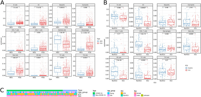
Figure 3.
Comparison of immune cell infiltration based on RNA-seq data by using Immune Infiltration module. (A) Comparison of immune infiltration between SARS-CoV-2-positive and -negative samples in GSE152075 nasopharyngeal swabs. Most of the immune cells increased during infection. (B) The difference of immune infiltrating cells between the subjects entering and not entering ICU, using GSE157103 peripheral blood leukocyte samples. (C) The population characteristics of all subjects in the GSE157103 dataset. The age of samples was not significantly correlated with whether the samples were admitted to the ICU (chi-square test p-value = 0.087). However, compared to Figure S3 (Old vs. Young), the change in immune infiltration was in line with figure (B).

