
Fig. 1. Expression of NωV coat protein in N. benthamiana.
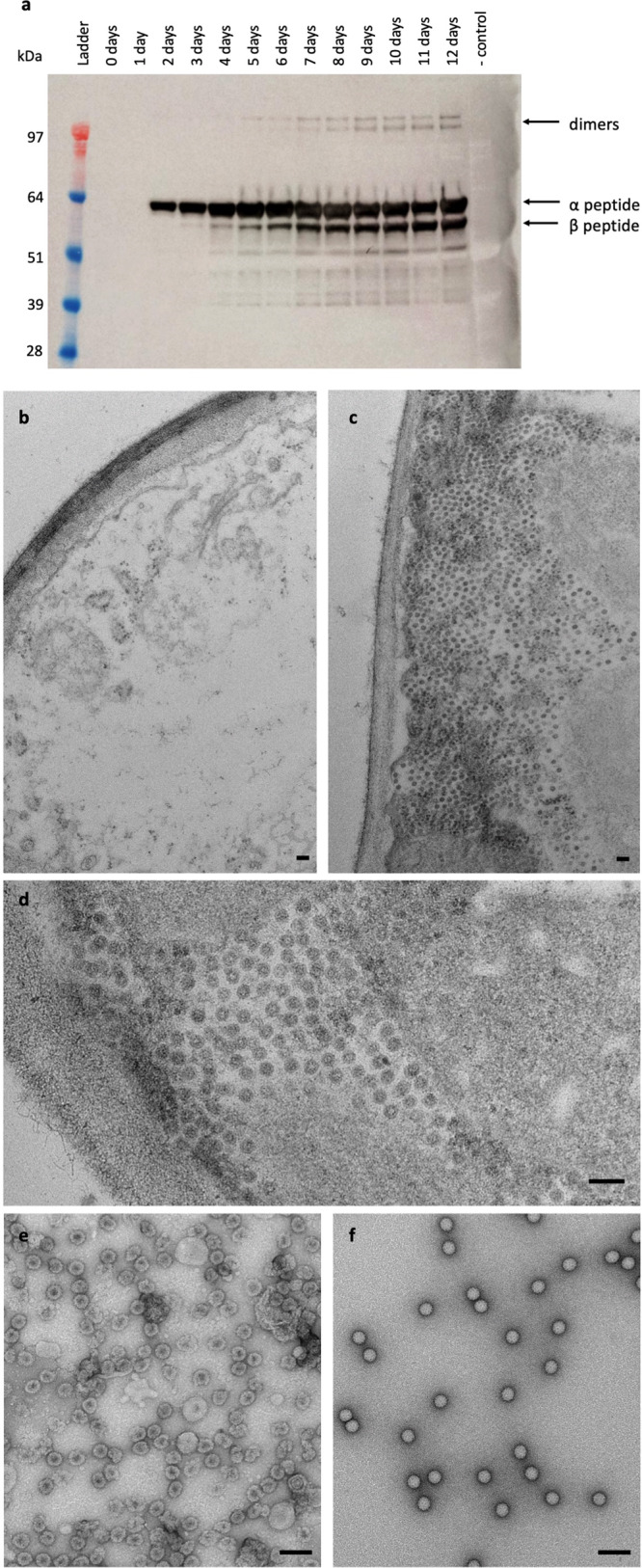
a Western blot of samples collected 0–12 dpi from leaves infiltrated with pEAQ-HT-NωV-WT. The negative control (- control) was from leaf material infiltrated with pEAQ-HT (empty vector). The protein was detected using a polyclonal antibody for the NωV coat protein. The positions of the uncleaved (α) and cleaved (β) versions of the coat protein are indicated, as are the positions of the dimeric forms. Ladder = SeeBlue Plus 2 pre-stained protein standards. Electron micrographs of thin sections of leaves 3 dpi with pEAQ-HT (b) or pEAQ-HT-NωV-WT at two different magnifications (c, d). All the leaf sections were counter-stained with 2% (w/v) uranyl acetate and 1% (w/v) lead citrate. Electron micrographs of purified procapsids (e) and capsids (f) negatively stained with 2% (w/v) uranyl acetate. For all micrographs the scale bar = 100 nm.