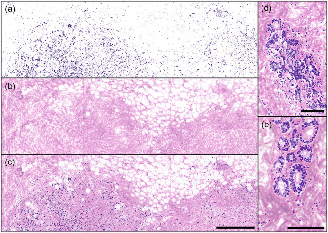
Fig. 4.
Individual and combined PARS absorption and scattering images of FFPE human breast tissues in block form, providing emulated H&E stain contrast. PARS images captured with sampling. (a) UV-PARS image of nuclei providing contrast analogous to hematoxylin staining of cell nuclei. The distribution of nuclei in the breast tissue specimen is evident but by itself is not diagnostic. (b) PARS scattering image providing visualizations analogous to eosin staining of cell membranes. This image reveals regions of adipose (clear voids) and dense, collagen-rich stromal tissue (pink-colored) and the organization of these structures in relation to each other. (c) Combined [(a) and (b)] emulated H&E PARS image of normal postmenopausal breast tissue with sparse and atrophic glands with higher nuclear densities (bottom left and right) with mainly adipose (top) and connective tissue (false-colored pink) comprising the majority of the sample. Scale bar: . Images are . (d), (e) Combined emulated H&E PARS images of normal postmenopausal breast tissue with atrophic glands surrounded by connective tissue. These smaller images are independent regions, not located within (a)–(c). (d) Scale bar: . Image is . (e) Scale bar: . Image is .

