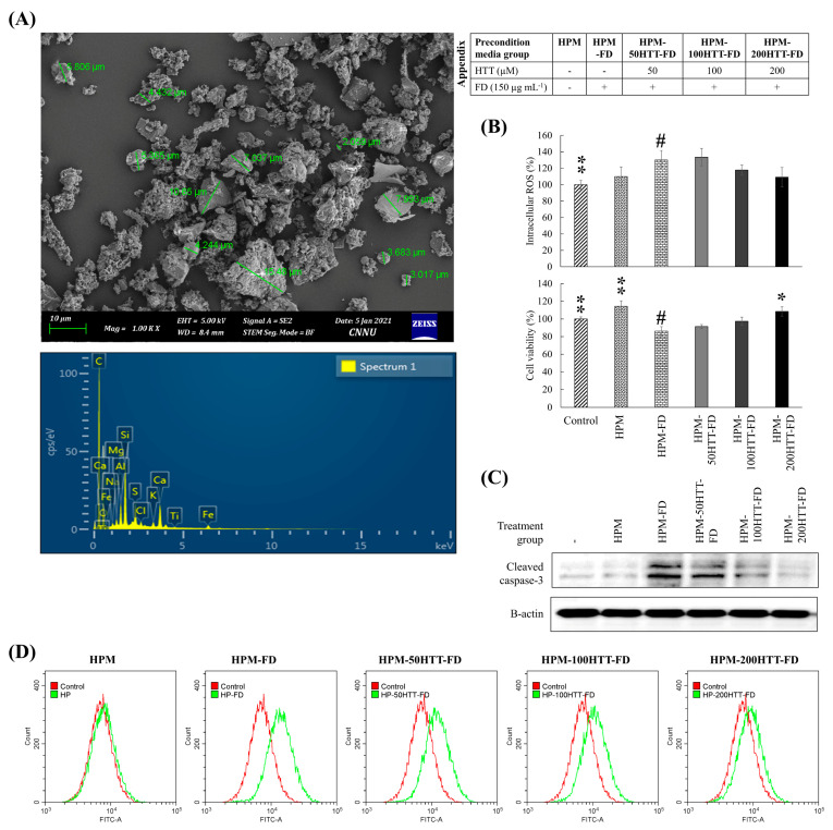
Figure 1.
Effect of HTT priming in FD-stimulated HaCaT keratinocyte preconditioned media on producing ROS in integrated HDFs and cytotoxicity. (A) SEM secondary electron image of the particle size distribution of FD and SEM-EDX analysis of FD. (B) Intracellular ROS level and viability of HDFs. (C) Levels of cleaved caspase-3. (D) Evaluation of intracellular ROS levels in HDFs by flow cytometry. HDFs were stimulated for 2 h with preconditioned media from FD-stimulated HaCaT keratinocytes with and without HTT pre-treatment. Intracellular ROS levels were measured 2 h after the stimulation period. Cell viability was evaluated after 24 h. Evaluations were carried out in triplicates (n = 3) and indicated as means ± SD. “*” and “**” respectively denote p < 0.05 and p < 0.01 if they are significantly different from those of HPM-FD treated HDFs “#”.

