Figure 5.
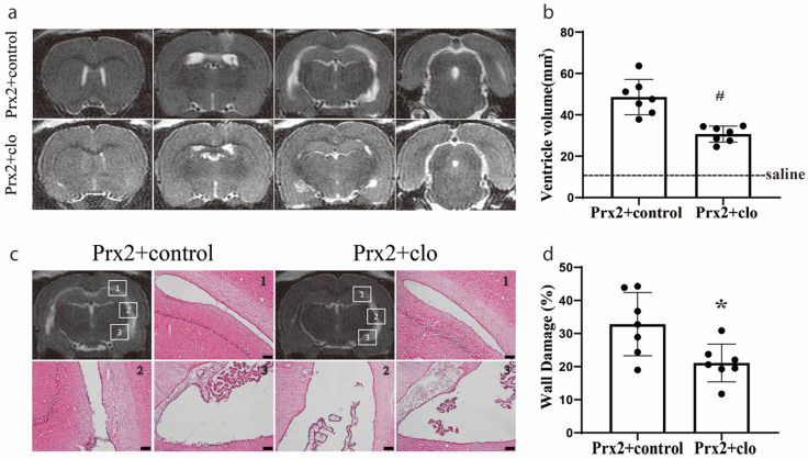
Injection of clodronate liposomes attenuated Prx2-induced hydrocephalus and ventricular wall damage. (a) Examples of T2 magnetic resonance imaging (MRI) in rats one day after injection of Prx2 + control liposomes (Prx2 + control) and Prx2 + clodronate liposomes (Prx2 + clo) into the right lateral ventricle. (b) Quantification of the ventricular volume in the two groups. Values are the mean ± SD; n = 7; # p < 0.01 vs. control group. (c) Examples of hematoxylin and eosin (HE)-stained sections showing ventricular wall damage one day after icv injection of Prx2 + control liposomes and Prx2 + clodronate liposomes. (d) The percentage of ventricle wall that was damaged is quantified in the bar graph. Values are the mean ± SD; n = 7; * p < 0.05 vs. control groups. Scale bar = 100 μm. Dots represent data for each animal.

