Figure 2.
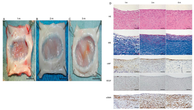
Data originally from Ichihara et al., 2015 demonstrating the macroscopic and histologic evaluation of a tissue-engineered arterial patch (TEAP) [78]. Macroscopic findings of each explanted tissue-engineered arterial patch at 1 month (A), 3 months (B) and 6 months (C) after implantation, showing no intimal hyperplasia and no aneurysm formation. No thrombus was observed. All inner sides of the patch were covered with native intima-like tissue with a smooth surface. Scale bar, 10 mm. (D) Hematoxylin–eosin (HE) and Masson staining (MS) at each observation end-point of bioabsorbable aortic patch. White and blank spaces (residual sponge polymer, arrowhead), which were observed in the TEAP at 1 month, were occupied by cellular components (red) and extracellular matrices (blue) in MS at 3 and 6 months after implantation. These findings showed good cell and tissue proliferation in the TEAP over time. Immunohistochemical staining for vWF, VEGF and αSMA (lower) is also shown. The luminal surfaces of the TEAPs were covered with a single layer of endothelial cells stained with antibodies to vWF at 1 month. The vWF-positive cells accumulated more clearly on the luminal surfaces of TEAP at 6 months. VEGF-positive cells in the patches were observed more on both the luminal surfaces and within the media 1 month after implantation; however, no VEGF expression was detected at 3 and 6 months. αSMA-positive cells were observed in the media of the regenerated tissue, and gradually increased over time. Original magnification HE, MS, vWF and αSMA ×200, VEGF ×100; scale bar, 100 μm. HE: haematoxylin–eosin; MS: Masson staining; TEAPs: tissue-engineered arterial patches; vWF: von Willebrand factor; VEGF: vascular endothelial growth factor; αSMA: alpha-smooth muscle actin.

