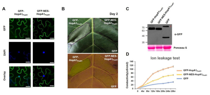
Figure 3.
The nuclear pool of HopA1Pss61 is required for ETI responses. (A) GFP-HopA1Pss61 and GFP-NES-HopA1Pss61 constructs were transiently expressed in N. benthamiana leaves using Agrobacterium adjusted to an OD600 of 0.2. Two days after infiltration, leaf discs were stained with 4′,6-diamidino-2-phenylindole (DAPI) for nucleus detection. Two days after infiltration, cells expressing fusion proteins were analyzed by an Olympus fluoview FV1000 confocal microscope under GFP fluorescence (top), DAPI (middle), and GFP/DAPI overlay (bottom). Scale bar: 20 μm. (B) GFP-hopA1Pss61, GFP-NES-HopA1Pss61, and GFP constructs were transiently expressed in N. tabacum cv. Xanthi leaves using Agrobacterium adjusted to an OD600 of 0.2. The photographs were taken under visible light (top) and UV light (bottom) at 2 days post-inoculation; (C) Detection of HopA1 protein by Western blot. Expression of GFP-HopA1Pss61, GFP-NES-HopA1Pss61, GFP in samples shown in (A) and (B) was confirmed by Western blot with anti-GFP antibody. Total protein was extracted from six leaf discs at 2 days post-inoculation. Total protein staining (Ponceau S) confirmed equal loading in Western blot analysis. The asterisk indicates a non-specific band cross-reacting with the anti-GFP antibody; (D) Quantification of cell death triggered by the hopA1 constructs described in (B) using electrolyte leakage. Error bars indicate standard deviation. Conductivity was measured at the indicated time points. This experiment was repeated twice with a similar result.

