Figure 1.
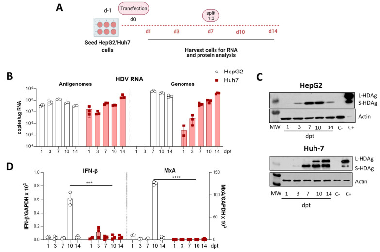
The HDV life cycle is supported in Huh-7 cells upon transfecting HDV-encoding plasmid but not in HepG2 cells. (A) Schematic representation of the experimental layout. HepG2 or Huh-7 cells were seeded and transfected with equal amounts of the plasmid encoding the HDV antigenome. For RNA and protein analysis, cells were collected at 1-, 3-, 7-, 10- and 14-days post-transfection (dpt), and cells were split 1:3 at 7-dpt. (B) Total RNA was extracted from cells and HDV antigenome and genome levels were assessed by RT-qPCR. (C) Western Blot analysis of HepG2 and Huh-7 cell lysates was performed to detect S-HDAg and L-HDAg. Positive control (C+): Huh-7 cells transfected with plasmids expressing S-HDAg and L-HDAg antigens and collected at 3 dpt. Negative control (C-): non transfected Huh-7 or HepG2 cells. (D) IFN-β (left) and MxA (right) expression levels were quantified by RT-qPCR and normalized using GAPDH as housekeeping gene. Statistical analysis using Mann–Whitney test revealed differences between the two cell lines (*** p < 0.001, **** p < 0.0001).

