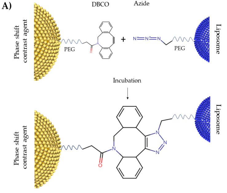
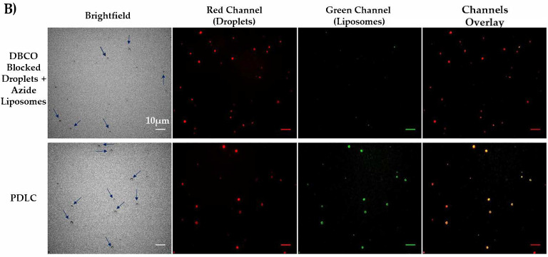
Figure 2.
(A) Schematic figure for DBCO-Azide click chemistry. (B) Fluorescent Microscopy of PDLCs after washing the unbound liposomes. Phase changeable droplets are dyed with DiD (red) and liposomes are dyed with DiO (green). There are no liposomes bound to the droplets in the top row since DBCO binding groups on droplets’ shell in this sample are blocked with sodium azide. Colocalization of green liposomes and red droplets (yellow) in the PDLC sample shows the clustering of droplets and liposomes. All scale bars show 10 µm.


