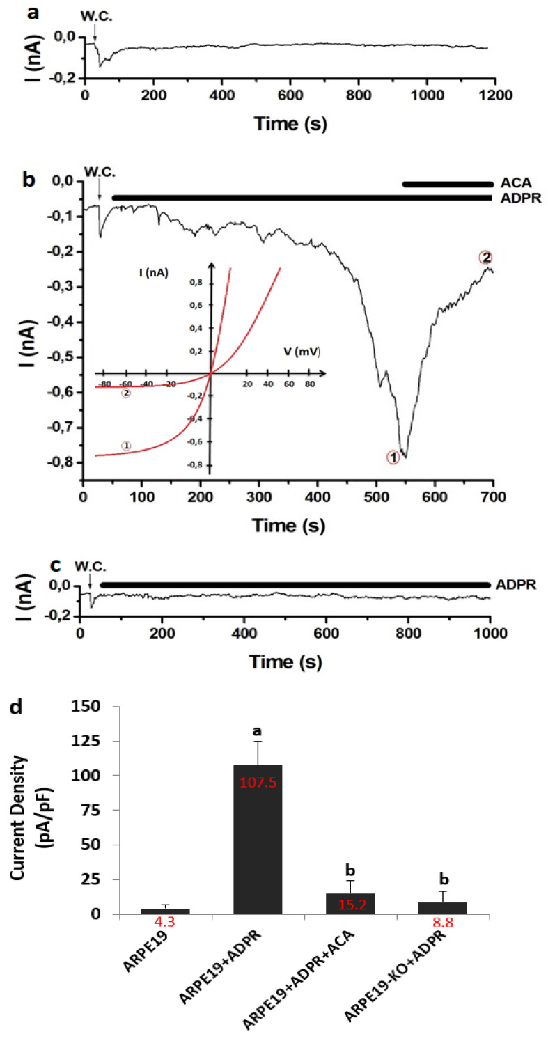
Figure 5.
The stimulation of ADPR induced the activation of TRPM2 in the cells of ARPE19, but not in the cells of ARPE19-KO. (Mean ± SD and n = 6). The whole cell (W.C.) configuration current records of TRPM2 were taken in voltage-clamp (at −60 mV) (a) The cells of ARPE19 without cytosolic ADPR (1 mM). (b) ARPE19 + ADPR group. The cytosolic ADPR (1 mM)-mediated TRPM2 currents were inhibited by ACA (25 µM). (b)-I/V. Time points ADPR and ACA were indicated 1 and 2, respectively. (c) ARPE19-KO + ADPR group. There is no cytosolic ADPR (1 mM)-mediated TRPM2 current. (d) The mean current densities from the cells of ARPE19 and ARPE19-KO. (a p ≤ 0.05 vs. ARPE19). b p ≤ 0.05 vs. ARPE19 + ADPR).

