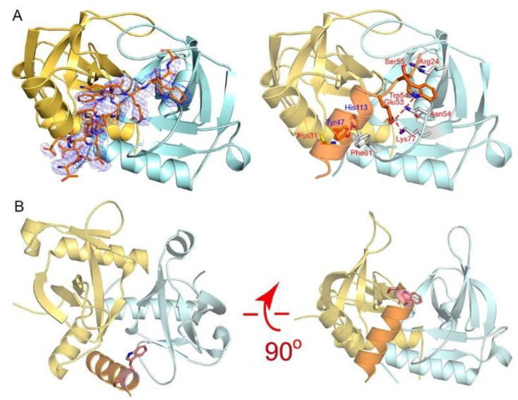
Figure 4.
The recognition mechanism of the MazE-mt1 (α3) peptide by MazF-mt1. (A) The cocrystal structure of the MazE-mt1 (α3)/MazF-mt1 complex (PDB 7DU5) and detailed interactions are shown on the right. The electron density of the OMIT map is shown by the blue mesh and was contoured at 2σ. (B) The structure of the MazEF-mt1 complex with only the α3-peptide of the antitoxin being shown. Two orthogonal views of the latter are shown, and the Trp54Ser55 dipeptide is shown in sticks. Note that the view on the right was the same as that of Figure 4A.

