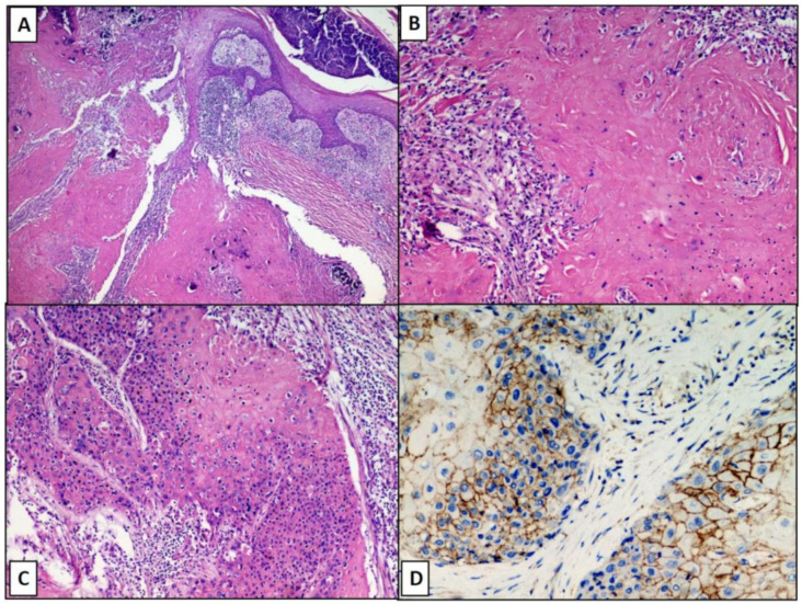
Figure 2.
Pilomatrixcarcinoma appears as a poorly circumscribed and asymmetric neoplasm, without ulceration of overlying epidermis and with necrotic areas (original magnification 10×; A); Detail of necrotic areas with the focal presence of shadow cells (original magnificaton 20×; B); basaloid cells surrounding the areas with necrosis en masse (original magnification 20×, C); predominantly membrane positivity (but also weakly cytoplasmic) for Beta-catenin at the level of matrical cells (IHC, Beta-catenin antibody, 40×, D).

