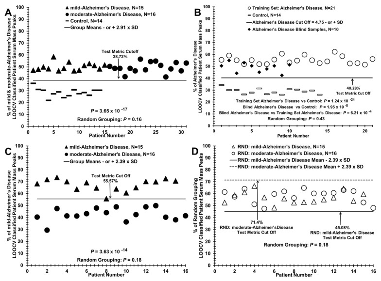
Figure 3.
Assigning pathology grouping to a blinded set of AD patient sera vs. controls and discriminating sera of mild AD patients from moderate AD patients. (A) Distinguishing mild and moderate AD patient sera as a group (N = 31, dark triangles and circles) versus controls (N = 14, dashes) for formation of the training set used in panel B. (B) Assigning correct group pathology to a blinded “left out” group of AD patients (N = 10, dark diamonds) from panel A against a training set of AD patients, N = 21 (open circles) and controls N = 14 (dashes) from panel A. (C) Distinguishing mild AD patient sera (dark triangles) from moderate AD patient sera (dark circles) using the LOOCV/PCV procedure. (D) Randomization of the two groups in panel C showing non-discrimination.

