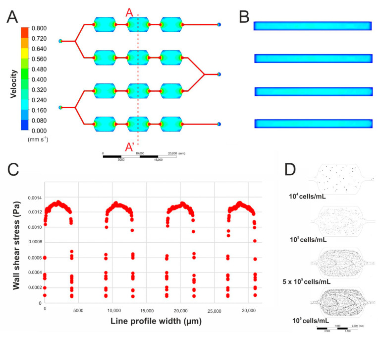
Figure 3.
(A) A simulation of the culture medium flow in the designed microfluidic system. (B) A simulation of the culture medium flow in the culture microchambers (cross-section view). (C) Wall shear stress simulation in four culture microchambers. (D) Simulations of cell distribution after introducing a suspension of cells of different densities into the microsystem.

