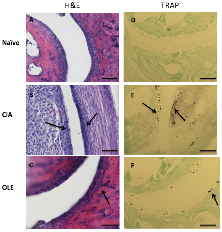
Figure 2.
Histological representative images of frontal joint section at Day 43: effects of dietary OLE (0.025% w/w) in synovial tissue, cartilage damage and bone erosion. Paw section from mice (n = 10) was stained with H&E (A–C) to show inflammatory activity and TRAP (D–F) staining for bone erosion. (A,D) Naïve group, non-arthritic control animals fed with SD; (B,E) CIA, induced arthritic animals fed with SD; (C,F) OLE, induced arthritic animals fed with OLE enriched diet. Joint cartilage thinning due to arthritic damage (asterisk) and inflammatory infiltrates (arrow) were observed in SD-CIA mice (B), as well as increased osteoclast cells (arrow) (E). Original magnification ×100 (A–C) and ×200 (D–F). The pictures are representative of at least six independent experiments with similar results.

