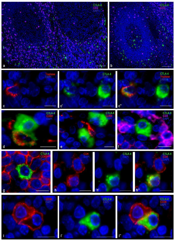
Figure 1.
Histoarchitectonics and immunophenotype of CTLA-4(+) tonsillar cells stained with antibodies of the SP355 clone. (a) Predominant localization of CTLA-4(+) in the extrafollicular regions of the tonsil. (b) Location of CTLA-4(+) cells in the germinal center of the lymphatic follicle and in the perifollicular zone. (c–c″) Cells with moderate (tryptase) and high levels of CTLA-4 expression. (d) Two CTLA-4(+) cells, one of which expresses CD68. (e) CTLA-4(+) cells are in contact with type 1 macrophage (presumably). (f) CTLA-4(+) cells contacting CD3+, lack of co-expression. (g) CTLA-4(+) cell surrounded by CD4+ cells. (h–h″) Co-expression of CD4 and CTLA-4. (i–i″) Co-localization of CTLA-4 and CD38 (plasma cell). Scale bar 125 µm for (a,b), and 10 µm for the rest.

