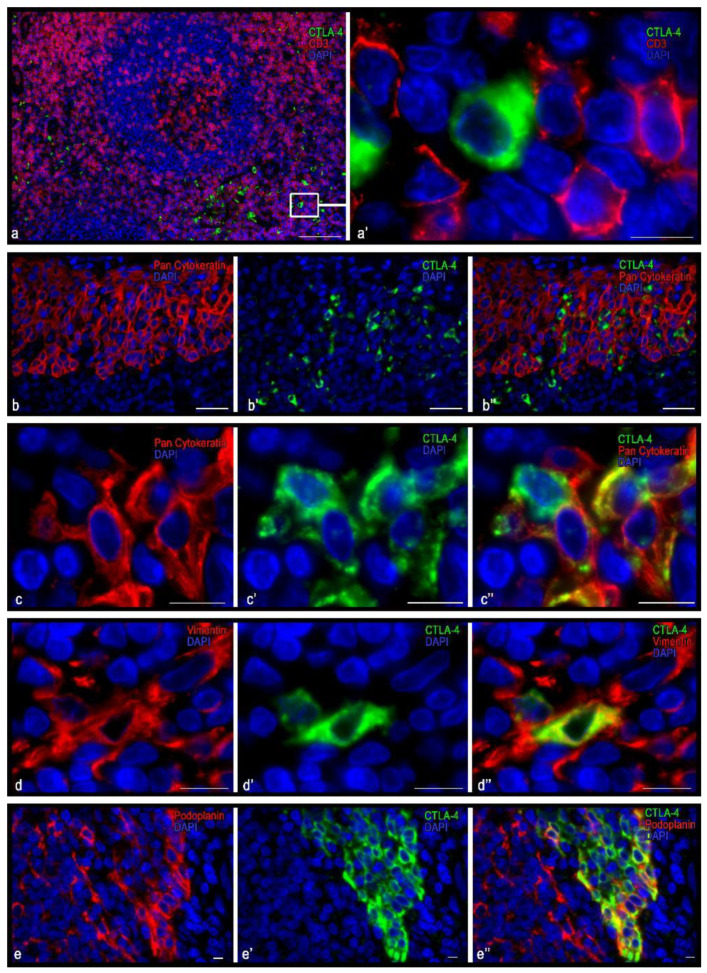
Figure 3.
Histoarchitectonics and immunophenotype of CTLA-4(+) tonsillar cells when stained with antibodies of the UMAB249 clone. (a) Localization of CTLA-4(+) cells in interfollicular lymphoid cords. (a′) An enlarged portion of figure (a). CTLA-4(+) cells do not express CD3 but are adjacent to and in contact with lymphocytes. (b–b″,c–c″) CTLA-4(+) cells in the reticular epithelium at low (b) and high (c) magnification. Obviously, some CTLA-4(+) cells express cytokeratins, while others do not. (d–d″) Immunopositive cells for vimentin, two of which express CTLA-4. (e–e″) The area of the reticular epithelium, protruding into the own plate of tonsils. Epithelial cells have an intense expression of CTLA-4. Moreover, the basal layers of the epithelium, as well as some cells of the more superficial layers, have immunopositivity for podoplanin. Scale bar is 125 µm (a), 50 µm (b), and 10 µm (a′, and the rest).

