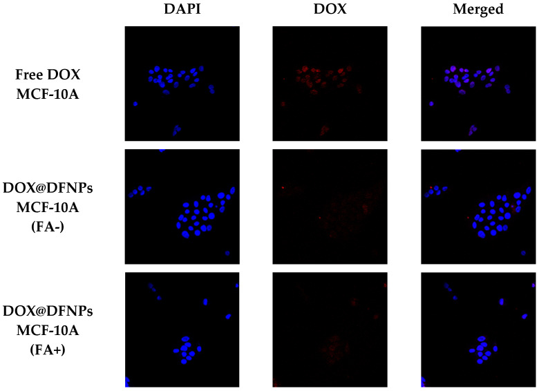
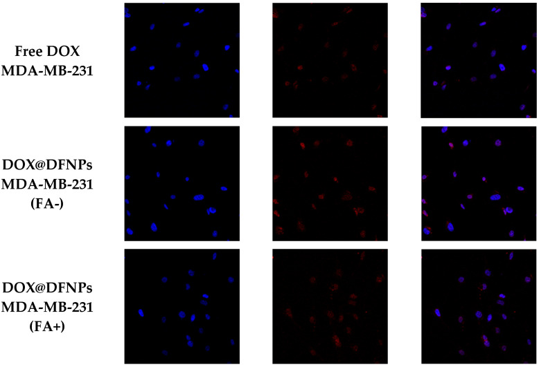
Figure 10.
Confocal fluorescence images (40×) showing intracellular DOX or DOX@DFNPs (red fluorescence) and nuclear DAPI (blue signal) in MDA-MB-231 and MCF-10A cells. In (FA+) images, FA receptors were blocked by 1 h pretreatment with free FA, whereas (FA-) images were acquired in the absence of this receptor ligand.


