. 2019 Dec 19;11(3):631–642. doi: 10.1039/c9sc05043d

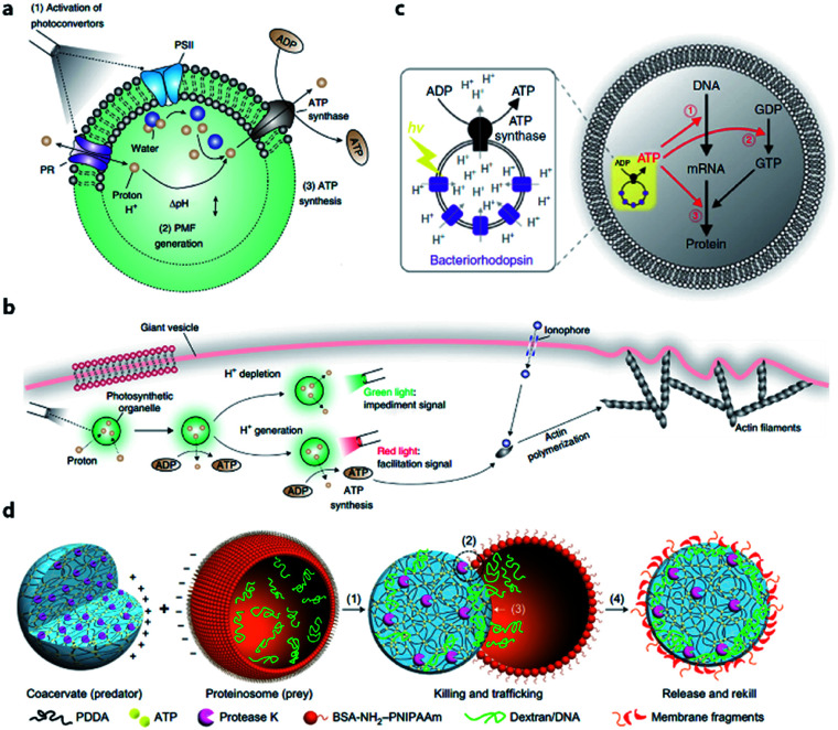
Fig. 3. (a) Structure and function of an ATP-generating artificial organelle with two photoconverters, plant-derived photosystem II (PSII) and bacteria-derived proteorhodopsin (PR), and an ATP synthase integrated into the membrane. PSII can be activated by red light to generate protons inside the organelle and PR can be activated by green light to deplete protons. The proton gradient across the organelle membrane drives the conversion of ADP to ATP by ATP synthase. PMF, proton motive force. (b) ATP-generating artificial organelles are encapsulated in a protocell. The synthesized ATP fuels ATP-dependent actin polymerization, thus inducing a morphological change in the protocell. Reproduced from ref. 53 with permission. Copyright 2018 Springer Nature. (c) Schematic of a protocell encapsulating an artificial photosynthetic organelle equipped with bacteriorhodopsin (bR) and F0F1-ATP synthase. Synthesized ATP is consumed for (1) mRNA transcription, (2) phosphorylation of guanosine diphosphate (GDP), and (3) aminoacylation of tRNA. Reproduced from ref. 54 with permission. Copyright 2019 Springer Nature. (d) Schematic representation of a synthetic protocell community including predator and prey. A protease K-containing coacervate microdroplet acts as an artificial predator protocell, which can capture the proteinosome-based prey through four steps: (1) electrostatic attachment; (2) protease-induced disassembly; (3) payload transfer; and (4) release of the compositionally modified predator protocell. Reproduced from ref. 57 with permission. Copyright 2017 Springer Nature.

Fig. 3