
Figure 1.
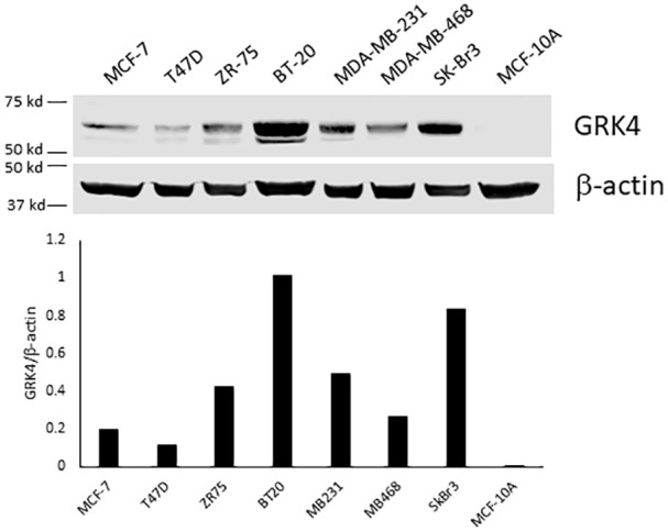
Western blot analysis of GRK4 in breast cancer and benign mammary epithelial cell lines. The cells were grown to confluence in serum containing medium without any treatment. Cell lysate containing 50 µg protein was separated on 10% SDS polyacrylamide gel and then transferred to a nitrocellulose membrane. The membrane was probed with monoclonal anti-GRK4 antibody (D-11). The experiment has been repeated 3 times with different GRK4 antibodies with similar results. Shown here is a representative blot.