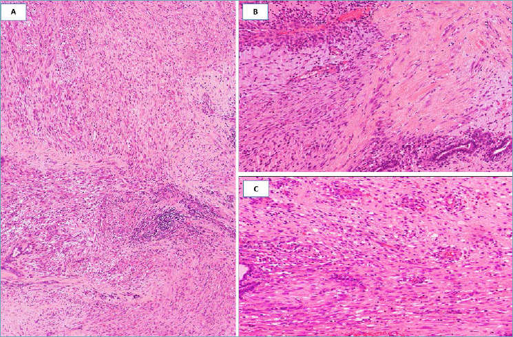
Fig. 2.

Nodular fasciitis. (A) Spindle cell proliferation with fibrous stroma and entrapped mammary ducts at the periphery of the lesion; (B) area with myxo-edematous stroma containing inflammatory cells (tissue culture-like appearance); (C) extravasated erythrocytes can be seen.
