
Fig. 5.
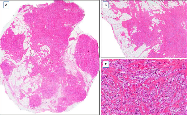
Lipomatous myofibroblastoma. (A) Fibrolipomatous tumor with pushing borders; (B) the fibrous component exhibits a finger-like infiltration into the lipomatous component, but the margins are circumscribed; (C) tumor area with the characteristics of classic-type myofibroblastoma: fascicles of spindle cells separated by keloid-like collagen bands.