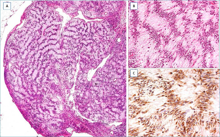
Fig. 6.

Palisaded/Schwannoma-like Myofibroblastoma. (A) Tumor with pushing borders, closely reminiscent of Schwannoma; (B) higher magnification showing nuclear palisading with formation of Verocay-like bodies; (C) cells, negative to S100 protein, are stained with α-smooth muscle actin, revealing their myofibroblastic nature.
