
Fig. 16.
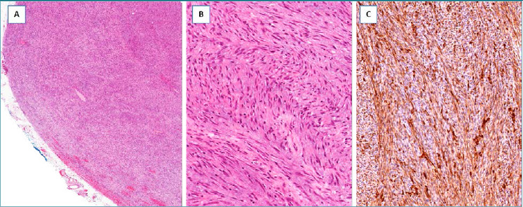
Low-grade myofibroblastic sarcoma. (A) Low-magnification showing a hypercellular tumor with pushing borders; (B) the neoplastic cells, with the morphological features of myofibroblasts, are arranged in short intersecting fascicles; (C) neoplastic cells are diffusely stained with α-smooth muscle actin.