Figure 6.
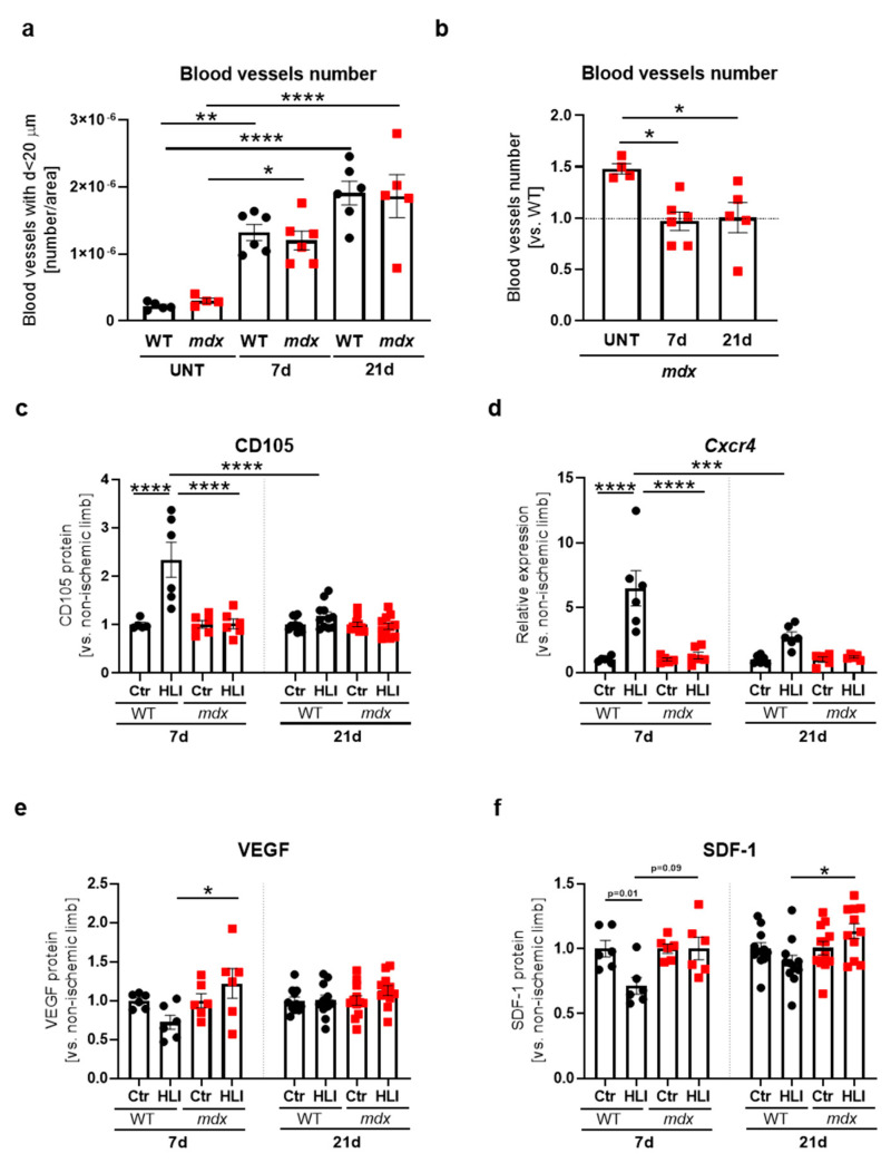
Blood vessels and angiogenic markers in the gastrocnemius of 12-week-old WT and dystrophic mice following hindlimb ischemia (HLI). (a) The number of blood vessels with a diameter lower than 20 µm in untreated (UNT) WT and mdx mice as well as in animals 7 and 21 days after HLI. (b) The total number of blood vessels in mdx mice shown in relation to WT mice after HLI; quantification based on immunofluorescent staining of CD31+α-SMA+ double-positive blood vessels; n = 4–6/group. (c) The protein level of CD105; ELISA, n = 5–11/group and (d) Cxcr4 transcript; qRT-PCR, n = 5–6/group. (e) Vascular endothelial growth factor (VEGF); ELISA, n = 6–12/group and (f) stromal derived factor-1 (SDF-1); ELISA; n = 6–12/group in the gastrocnemius of WT and mdx mice 7 and 21 days after HLI. The results are presented as mean ± SEM. * p < 0.05, ** p < 0.01, *** p < 0.005, **** p < 0.0001 by one-way ANOVA with Tukey’s posthoc test.

