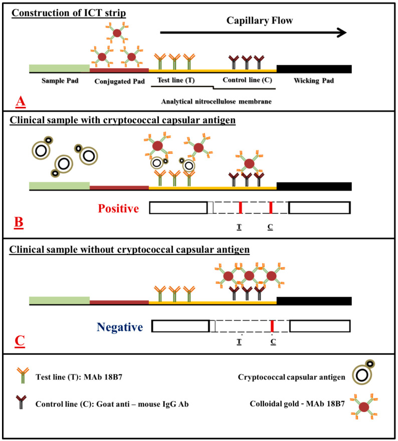
Figure 1.
A schematic of mAb 18B7 sandwich ICT strip for the rapid detection of cryptococcal capsular antigen from clinical samples. (A), the schematic presentation of the positions where colloidal gold conjugated mAb 18B7, mAb 18B7 and goat anti-mouse IgG antibodies are immobilized in an analytical nitrocellulose membrane. (B), the reactions that occur on the mAb 18B7 sandwich ICT strip in the presence of cryptococcal capsular antigen and (C), in the absence of cryptococcal capsular antigens. The red–purple color of colloidal gold conjugate appears at the test line and/or control line, depending on the presence or absence of cryptococcal capsular antigen in the clinical sample.

