Figure 1.
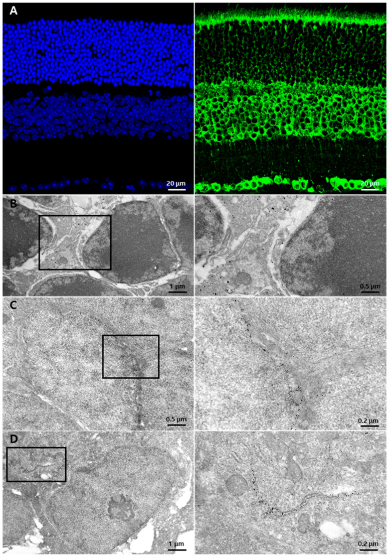
GRP78 expression in normal retinas: (A) Representative normal retina labeled with DAPI (blue) and GRP78 (green). GRP78 was mainly labeled in the rod and cone layers and the cell bodies of the INL and GCL. In the ONL, the photoreceptor cell bodies were weakly surrounded by GRP78. (B–D) Representative EM images of the normal retina with immuno-gold-labeled GRP78. (B) Normal photoreceptor cells in ONL. Immuno-gold labeled-GRP78 was detected in the interspace of the photoreceptor cell bodies but not in the photoreceptor cells. (C) Cell bodies in INL. Immuno-gold-labeled GRP78 was detected in the perinuclear spaces of INL cells. (D) Representative EM images of GCL. Immuno-gold-labeled GRP78 was detected in the perinuclear spaces and ER lumen of the GC. INL, inner nuclear layer; GCL, ganglion cell layer; ONL, outer nuclear layer; EM, electron microscopy; ER, endoplasmic reticulum; GC, ganglion cell.

