Figure 2.
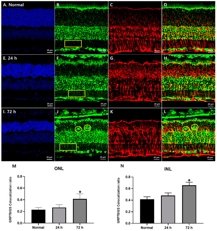
GRP78 expression in Müller glial cells in blue LED-induced RD retinas. (A,E,I) DAPI (blue) stained retina sections of the normal (A), 24 h (E) and 72 h (I) retinas after blue LED exposure. ONL thickness prominently decreased at 72 h. (B,F,J) GRP78 (green)-labeled sections of normal (B), 24 h (F) and 72 h (J) retinas after blue LED exposure. GRP78 was increased in the ONL and IPL in a time-dependent manner after LED exposure (yellow rectangles) and labeled in giant cells in the ONL (yellow circles). (C,G,K) GS (red)-labeled retina sections of the normal (C), 24 h (G) and 72 h (K) retinas after blue LED exposure. GS-labeled Müller cell processes enlarged and encircled photoreceptor cells in the ONL after injury. (D,H,L) Merged results of GRP78 and GS. GRP78 was increased in the Müller cell processes from IPL to ONL in a time-dependent manner while GRP78-labeled giant cells in the OPL showed no GS immunoreactivity (yellow circles). (M,N) Co-localization ratio of the GRP78/GS in the ONL (M) and IPL (N). In both the ONL and INL, the GRP78 co-localization ratio in GS-labeled Müller glial cells was significantly increased at 72 h after LED exposure (n = 7, p < 0.05 (*), ANOVA).

