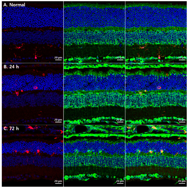
Figure 3.
GRP78 expression in microglial cells in blue LED-induced RD retinas. (A) A representative confocal image of the normal retina labeled by DAPI (blue), GRP78 (green) and IBA1 (red). IBA1-labeled microglial cells were weakly labeled by GRP78 in the normal retina. (B). After 24 h of light exposure, microglial cells were detected in the ONL with an enlarged shape and increased GRP78 compared with the normal retina. (C) After 72 h of light exposure, microglial cells were markedly increased in the ONL and GRP78 was prominently increased in microglial cells compared with those after 24 h.

