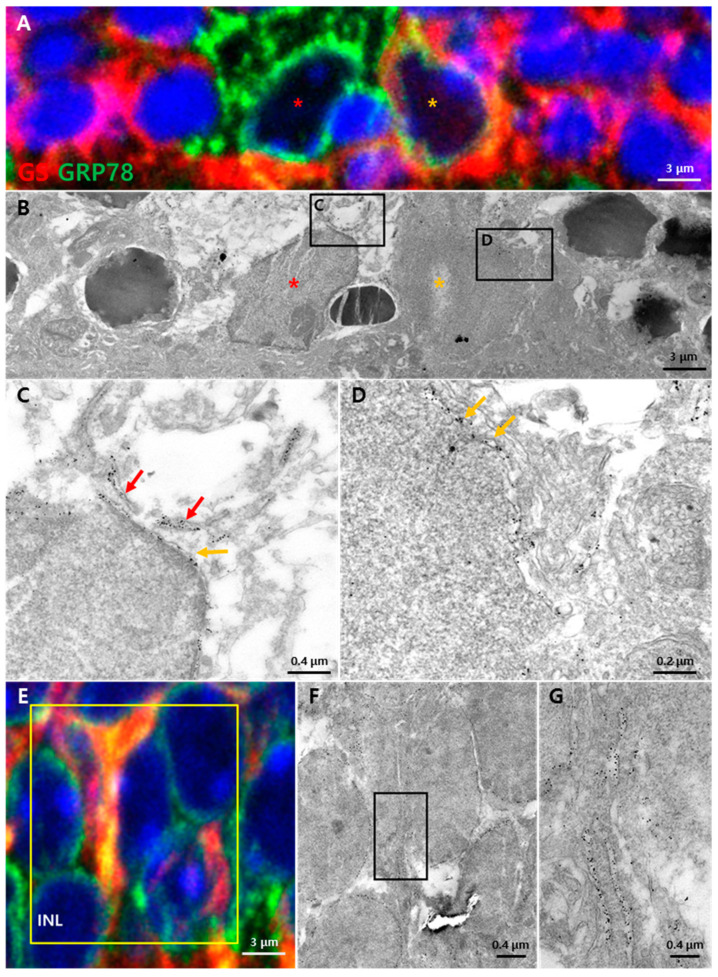
Figure 4.
Subcellular GRP78 localization in the injured retina 72 h after light exposure. Semi-thin sections of the retina stained by GRP78 (green, gold), GS (red) and DAPI (blue). (A) Microglia and migrated Müller glia in the ONL. Microglia (red asterisk) were more strongly labeled by GRP78 than Müller glia without GS while Müller glia (orange asterisk) were labeled by both GRP78 and GS. (B) EM images of microglia (red asterisk) and migrated Müller glia (orange asterisk) in the same region as (A). (C) Immuno-gold-labeled GRP78 was detected in the perinuclear space (orange arrow) and lumen of the ER structure (red arrows) of the microglia. (D) Immuno-gold-labeled GRP78 was detected in the perinuclear space of the Müller glial cell (orange arrows). (E) Confocal images in the INL of an injured retina with Müller glia processes. (F) EM images matched with the yellow box in (E). Processes of the Müller glia in the INL labeled by GRP78. (G) A higher magnification of the Müller glia process in (F). Membranous structures containing immuno-gold-labeled GRP78 located in Müller glia processes.

