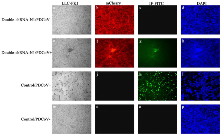
Figure 4.
Effects of pSil-double-shRNA-N1-mcherry plasmids on PDCoV-induced CPE in LLC-PK1 cells by immunofluorescence assay. (a–d) pSil-double-shRNA-N1-mCherry stably transfected cells served as a mock transfection control; (e–h) pSil-double-shRNA-N1-mCherry stably transfected cells indicated a noticeable reduction of PDCoV N protein expression; (i–l) non-transfected cells indicated no obvious effects on PDCoV infection as a positive control; (m–p) non-transfected cells without PDCoV infection were used as a negative control. The green and red colors show the presence of PDCoV N protein-positive cells and stably shRNA-transfected cells, respectively. The nuclei of the cells were stained in blue by DAPI.

